The Cardiovascular, Blood, and Lymphatic Systems
Blood
Blood is one component of the cardiovascular system, the others being the heart and blood vessels. As the body’s transport medium, the blood provides a means to transport oxygen, carbon dioxide, hormones, nutrients, and waste. The blood also contains most of the cells, proteins, and chemicals required to initiate and support resistance to diseases. Body temperature and pH are also regulated by the blood.
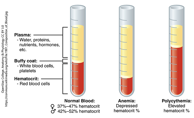
Blood is composed of plasma (55%) and formed elements (45%). Plasma is the straw colored fluid that is made up of water, salts, and proteins. The formed elements consist of the red blood cells (erythrocytes), the white blood cells (leukocytes), and the platelets (thrombocytes). Red blood cells contain a protein called hemoglobin that helps transport oxygen in blood and is responsible for blood’s bright red color. White blood cells help fight infection and consist of:
- neutrophils,
- eosinophils
- basophils
- monocytes
- lymphocytes
Platelets are responsible for clotting blood.
Lymphatic System
The lymphatic system is a system that runs throughout the body and is made up of lymphatic vessels and supporting organs that drain excess interstitial fluid, transport dietary lipids, and facilitate immune responses. The lymphatic system includes:
- lymphatic fluid (lymph)
- lymphatic capillaries
- lymph vessels
- trunks
- ducts
- lymph nodes
- red bone marrow
- thymus gland
- spleen
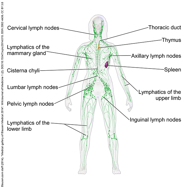
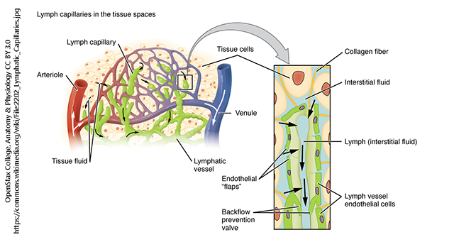
Lymph fluid comes from the blood and contains white blood cells. As oxygen and nutrients are diffused at the capillary level to all cells, lymph capillaries are also present. As fluid goes between cells the blood becomes interstitial fluid and then either enters back into the bloodstream or into the lymph capillaries. Some fluid diffuses into the lymph capillaries and then into the lymph vessels. The lymph vessels transport lymph to the lymph nodes to be filtered. Lymph nodes are clusters of lymph tissue in the neck (cervical lymph nodes), armpit (axillary lymph nodes), and groin (inguinal lymph nodes). There are also clusters in the intestines called Peyer patches. After the lymph is filtered it is returned to the bloodstream through a duct in the chest.
Lymphocytes are produced in the bone marrow and matured in different parts of the body. B lymphocytes mature in the bone marrow and T lymphocytes mature in the thymus. Both B cells and T cells are part of the immune response and coordinate different ways the body attacks foreign invaders such as viruses and bacterias.
The tonsils are located at the back of the throat and contain white blood cells which help trap and kill germs. The spleen is located in the left upper quadrant and is also made of lymphatic tissue. It filters blood and removes old and damaged red blood cells.
Heart & Blood Vessels
The heart is a strong, muscular pump just a little over the size of a fist. The heart pumps about 3600 gallons of blood through the body in one day, beating ~100,000 times a day, about 39 million times per year. By the time you reach 65 years old, the heart has beaten two billion times. Each heartbeat ejects approximately 70 ml of blood into the systemic circulation.
The human heart is located within the thoracic cavity, medially between the lungs and the pleural membranes that cover them in the space known as the mediastinum. The heart is covered by a tough membrane known as the pericardium (pericardial sac) and sits in its own space called the pericardial cavity.
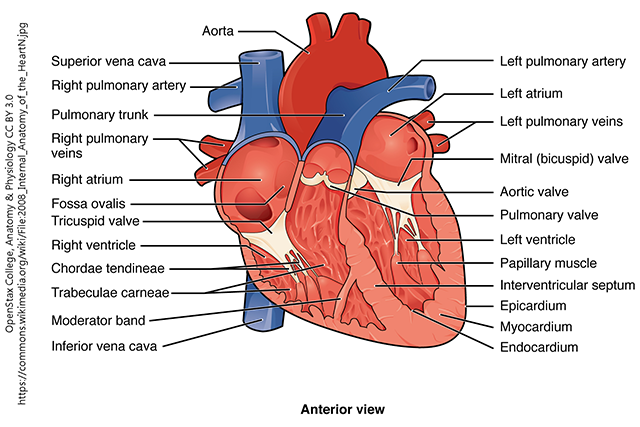
The human heart consists of four chambers: The left side and the right side are divided by a septum and each have one atrium and one ventricle. Each of the upper chambers, the right atrium (plural = atria) and the left atrium, acts as a receiving chamber and contracts to push blood into the lower chambers, the right ventricle and the left ventricle. The right atrium receives deoxygenated blood from the body; the left atrium receives oxygenated blood from the lungs. The ventricles serve as the primary pumping chambers of the heart, propelling blood to the lungs or to the rest of the body.
The heart has four valves. The atrioventricular valves control the flow of blood between the atria and ventricles. There are two atrioventricular (AV) valves in the heart: the tricuspid valve that is located between right atrium and right ventricle, and the bicuspid valve (mitral valve) that is located between left atrium and left ventricle. The semilunar valves control the blood leaving the heart from the ventricles. There are also two of these valves: the pulmonary valve is located between the right ventricle and pulmonary artery, and the aortic valve is located between the left atrium and aorta.
Circulation of the blood through the body is achieved through blood vessels. There are three main types of blood vessels: arties, veins, and capillaries. Arteries take blood away from the heart and carry oxygenated blood. The largest artery in the body is the aorta and the smallest arteries are called arterioles. Veins return blood back to the heart from the body. The largest vein is the vena cava. The superior vena cava returns blood from the arms and head, and the inferior vena cava returns blood from the trunk and legs. The smallest veins are called venules. Where arterioles and venules meet up in the body are called capillaries and this is where gas exchange occurs.
Circulation through the Heart – Summary
1. Blood from the superior vena cava and the inferior vena cava enters the right atrium
2. Blood flows from the right atrium to the right ventricle via the tricuspid valve
3. Blood is pumped from right ventricle to pulmonary artery (trunk) via the pulmonary valve
4. Blood is oxygenated in the pulmonary capillaries of the lungs
5. Blood returns to heart via pulmonary veins
6. Oxygenated blood from lungs returns to the left atrium
7. Blood flows through the mitral valve (also called the bicuspid valve) into the left ventricle
8. Blood is pumped through the aortic valve into the aorta
9. Blood distributed to body through branches of the aorta and other vessels into capillaries
10. Blood returns to heart via systemic veins and the venae cavae
The heart wall consists of three layers. The deepest layer is the endocardium which provides a smooth surface for blood as it flows through the chambers and the valves of the heart. The thick middle layer is the myocardium, the muscular wall of the heart. The muscular layer makes up 95% of the heart and is responsible for the pumping action. The thin, outermost layer of the heart is called the epicardium.
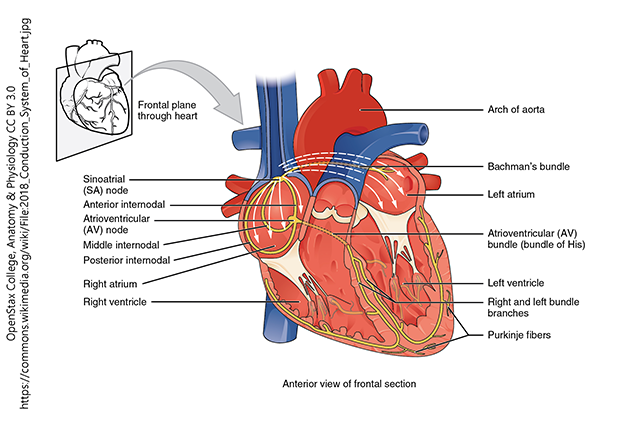
The heart has its own electrical conduction system that causes it to pump. The sinoatrial (SA) node controls the rhythm of the heartbeat and is known as the pacemaker of the heart. The SA sends an impulse to the atrioventricular (AV) node, and then travels to the bundle of His, then to the bundle branches, and finally to the Purkinje fibers causing the ventricles to contract. The heart muscle contraction is called systole (giving rise to the systolic blood pressure) and the relaxation is diastole (giving rise to the diastolic blood pressure). An electrocardiogram (ECG or EKG) is a recording of the electrical changes of the heart. Abnormal ECGs show problems within the conduction pathways of the heart. An ECG can show if the heart is enlarged, if certain regions of the heart are damaged, and the cause of chest pain.
Media Attributions
- Unit 8 figure 1 Composition of Blood © OpenStax College is licensed under a CC BY (Attribution) license
- Unit 8 figure 2 Lymphatic System Female © Bruce Blaus is licensed under a CC BY (Attribution) license
- Unit 8 figure 3 Lymphatic Capillaries © OpenStax College is licensed under a CC BY (Attribution) license
- Unit 8 figure 4 Internal Anatomy of the Heart © OpenStax College is licensed under a CC BY (Attribution) license
- Unit 8 figure 5 Conduction System of Heart © OpenStax College is licensed under a CC BY (Attribution) license
